 My major interests include developing the tools of Structural Biology, the pursuit of novel macromolecular structures and complexes, and their functional analysis. I employ single crystal x-ray crystallography and solution macromolecular small angle x-ray scattering (BioSAXS) to determine the structure and function of macromolecules and their complexes.
1) Crysta llography
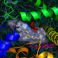
Protein crystallography can produce atomic resolution structures of macromolecules, probe oligomeric states, complexes, and even dynamics. The crystallographic study of proteins has resulted in several Nobel prizes, most recently for the calcium channel in 2003, the RNA polymerase in 2006, the ribosome in 2009, and the G-protein-coupled receptors in 2012. To speed structure discovery I am developing new computational crystallography and structure analysis tools. Some of these tools are available for download from the Protein Model Builder (PMB) web site. In my over two decades of experience and many collaborations I have studied areas ranging from protein folding, to oxidative stress response, cellular signaling, protein dynamics, antiviral molecules, DNA binding, RNA and DNA polymerases, and viral endocytosis. These studies have produced over 50 structures in the PDB and numerous publications.
2) Solution Small Angle X-ray Scattering of macromolecules (BioSAXS)
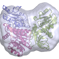
The solution x-ray scattering from macromolecules can yield information about more than just the average diameter (RG) and length of the molecule (DMAX). In addition the scattering data also encode information about the shape of the molecule, or molecular complex, and their distribution. In collaboration with many diverse groups we have used solution scattering to study molecular complexes, allosteric systems, amyloid formation, dynamic interactions, viral RNAs, mitochondrial polymerases, and cellular and neuronal signaling complexes. The molecules we examined have ranged in size from a few kDa to MDa complexes. As a consequence of these many collaborations I have developed several useful BioSAXS software utilities and web-based services which are made available on our SAXNS website.
3) The SCSB Macromolecular Structure X-Ray Laboratory
The SCSB x-ray facility offers the tools necessary to solve crystal structures and determine the solution structures of macromolecules and their complexes. Crystallization projects benefit from a series of instruments and facilities, including: a Malvern Zetasizer DLS, an ARI PHOENIX 96-well crystallization tray dispenser, a Rigaku Hotel/Minstrel tray imager, the Alchemist screen maker, and temperature controlled low-vibration environmental rooms. We offer two XRD systems: a Rigaku R-AXIS-IV with FR-E Superbright source and a Bruker D8-Venture with the Turbo TXS high-brilliance source. Our Rigaku BioSAXS-1000 camera coupled with the FRE source and automatic sample changer provide the optimal in-house SAXS system. The Structural Biology Computer Commons provides high-performance multi-CPU Linux workstations with 3D visualization for any structural project. For more information please visit the XRAY website.